
This page is to document the diagnosis and course of treatment of my
schwannoma. Comments and corrections, by physicists, physicians, etc.
are welcome. Please send them to my email
Disclaimers:
1) I am not a medical doctor, just a physicist that happened to get
"lucky" and found almost nothing about this on the web. The opinions
are my own, based on my research. They are only opinions.
2) Every case and every patient is different. What (I hope) is
working for me, may not be indicated for you. There is no substitute
for seeing a doctor, but this web site should at least help you go in
informed so that a fruitful discussion on options can ensue. I know
that in my case, my local Ear, Nose, & Throat doctor was discussing
possibilities that at the time were way over my head as I did not even
know what a schwannoma was or how serious it could become. The 2nd
discussion with him was much better as I had a handle on this.
Providing information to people who are as "lucky" as me, is really the
main purpose of this web site.
At this point in my understanding of how this is called, the doctors
seem to use Schwannoma and Neuroma interchangeably. The radiologist who
did the initial reading of the MRI labeled this as a schwannoma, but my
Ear Nose and Throat physician called it a Neuroma. The 7th
cranial nerve is also known as the facial nerve, thus the there are
also two names for the nerve. Clarification from the medical
community would be helpful.
I will put this description in terms closer to an electrical
engineer, instead of a doctor. Imagine your nerves as copper
wires. Around all wires that you are familiar with is an insulator to
protect and insulate the wire. Nerves have an analogous construction.
They too have an insulator surrounding the cells that carry the
signals. The Schwannoma is a benign (i.e. non-malignant or
non-cancerous) growth on these insulator cells. It is not a growth on
the nerve cells themselves. The growth is an extremely slow growth. I
am told it is about 1 mm per year.
Early in 2005, my left ear felt clogged up, like there was fluid in
the left ear. You know the feeling, you have it coming out of a
swimming pool or shower. The feeling of being clogged up did not go
away. Then while I was away in a hotel room, using my watch as an
alarm, my right ear was pressed against the pillow and my left ear was
free. I could not hear the watch alarm at all.
At this point I went to my general practitioner. He first checked to
see if there was any blockage on the external side of the ear
drum. There was not. He then performed a couple of simple tests that
showed conduction, not nerve hearing loss. He put me on a decongestant.
When that did not work, my GP punted and I went to an Ear Nose and
Throat specialist. At the ENT's office, more extensive testing was done
which showed that I had hearing loss on both the low frequency end
(which is attributed to conduction) and high frequency (which is
attributed to nerve damage). As he asked me questions about when and
how the hearing lost began, I apparently was not giving him "good"
answers, so he ordered an MRI. This brought out the real diagnosis.
At this point I started searching the web to find out where to go to
get this treated. I ended up at Stanford.
Next to the 7th nerve is the 8th or Acoustic nerve. Schwannoma's on
this nerve are much more common, and the treatment plan for the 7th
nerve is based on an assumption that the physiology of the Schwannoma's
are the same. The 7th nerve Schwannoma is so rare that the medical
community just does not have statistics on it.
A table of all of the Cranial nerves can be found here.
Here are 3 of the 232 images from the MRI. These have the contrast
agent
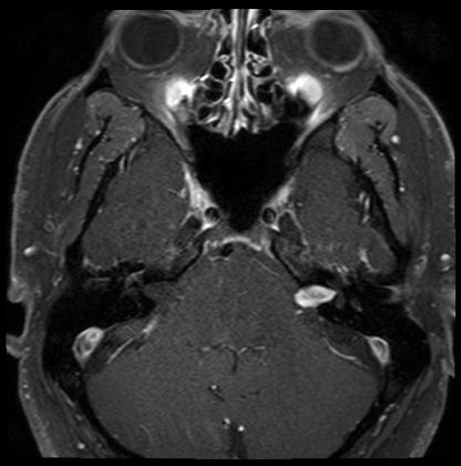
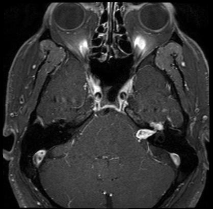
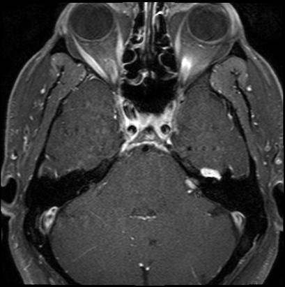
The Schwannoma is seen in light gray on a diagonal going from the
center of the image to the lower right.
Some sketches from the office of Dr. Jackler at the Stanford
Otolaryngology Clinic at Stanford University:
This sketch shows the Acoustic (Hearing), Balance and Facial nerve.
On the brain side of the hearing canal, they are all bunched up
together.
This is another sketch.
The radiologist's report says that the growth extends inwards to a
region called the Geniculate
Ganglion. I believe in talking with the doctors, that at the
present time, the growth does not hit this region. Continuing
with my simplified electrical engineer's view of the nervous system, if
the brain it self is the central processor, the Intel, AMD, or PowerPC
chip, the Geniculate Ganglion (GG) is either a smaller processor
attached to the brain where a number of the Cranial Nerves come in and
get processed / merged before going to the brain itself or if you know
what a ribbon cable is, it is the point that the ribbons come together
before going to the brain. I know that if the growth has not hit the
GG, then a splice can be done. I am not sure if a splice can be done
with today's technology if the growth has hit the GG itself.
Unfortunately there is no "good" course of action today. There is no
Star Trek doctor with a magic gizmo to waive over me and treat the
Schwannoma.
If I were in my 80's, this is probably what I would do. The growth
will happen slowly, leading to increased loss of hearing as it crushes
the neighboring 8th or Acoustic Nerve, as well as a palsy on the left
side of my face. In fact the palsy has begun with an intermittent
tremor in my lower left eyelid. Thinking about it, this tremor has been
with me longer than the loss of hearing, but I did not do anything
about it as I did not think it was symptomatic of anything major. After
the palsy, when the growth crushes and kills the facial nerve, then I
would get paralysis on the left side of my face. I don't know what
further complications would arise as the growth puts pressure on the GG
or on the brain itself. I also don't know if the growth could go on
inside the GG. But since I am not in my 80's and I know that the growth
is active (based on the recent loss of hearing), this is not an option.
Radiation, with either the Gamma Knife or Cyber Knife. In both
cases, beams of radiation are delivered along many different angles,
all intersecting at the desired target. Each beam by itself should do
minimal damage, but where they all intersect, the dose is now high
enough to damage the targeted areas.
To me, this course of treatment will be successful if the growth is
slowed down or even stopped. The best outcome would be an actual
shrinkage of the growth. It is possible that the treatment will fail
completely. It is also a finite possibility that 7 to 10 years down the
road the growth could turn malignant. There have been 8 cases cited in
the literature. These were all Acoustic Neuromas. All were fatal.
In the case of the Gamma Knife, this is done with Gamma Ray emission
from the decay of Cobalt 60. CO60's main decay path has a half life of
5.271 years. There are over 200 sources that are aimed along different
paths to intersect at the tumor. CO60 gives off 2 gamma rays when it
decays. One at an energy of 1.173 MeV and one at 1.332 MeV. Data
from here.
It
is
not clear to me if the Gamma Knife uses both or if it has a
filter to only use one. A description of the Gamma Knife can be found here
(on page 18) as well as here
or many other sources.
Based on what I understand of this technology, the head is bolted
into a frame and is not allowed to move at all. This apparently is very
uncomfortable, and as a result, they do not deliver the dose in
multiple sessions, but is done in one day. Delivering the dose is
multiple sessions is called "fractionation".
This is the first revision of this paragraph after my treatment now
that I know more.
There will be a medical physicist as part of the team that treats
me. I will be asking this person the questions I have outlined in
the above paragraph.
You can see a presentation by my Radiologist, Dr. Iris Gibbs, here.
In any case, this machine has advantages and disadvantages over the
Gamma Knife. Its main advantage to a patient is that there is no
bolting the head to an immovable frame. Day 1 of the treatment protocol
at Stanford is to put a mesh or grid over your head. This will mold to
your head. Then a CT Scan (cat scan) is done. Both the Schwannoma and
the grid are visible to the scan. Day 2 is an off day. Day 3 through 5,
you are put into a room with the CyberKnife, with the grid on your
head. The machine is continuously imaging the grid with soft x rays
(this is the one disadvantage) and dynamically adjusting for head
movement. The head is allowed to move up to 2mm. After that, the
software prevents the beam from firing.
I would think another advantage of this when the research is done,
is to see if there is a wavelength more harmful to the growth than to
either normal schwann cells (the insulating cells) or the nerve cells
themselves. Since the radiation is derived from a Linear Accelerator,
one has many more possibilities for output wavelength than one would
with a radioactive decay. Mother Nature only allows a describe and
finite set of wavelengths from decay.
This treatment option is not very attractive, and is a last resort.
Basically the area of the nerve under the Schwannoma is removed, and a
nerve from another part of the body is spliced in. For 12 to 24 months
there will be total paralysis on the left side of my face. During this
time, measures have to be taken to make sure that the left eyelids
blink to supply tears to the eye. This is done with some sort of
mechanical spring mechanism. Eventually healing does take place, and
some level of facial movement is restored. However the medical
community tells me that on a scale of 0 to 6, I will be at a
level 3 for facial movement. I don't know if this scale is linear or
not. I also will probably go totally deaf in the left ear.
The game plan therefore is to have some regular monitoring after the
CyberKnife procedure, and make sure that the growth is not getting too
close to the GG. I think based on my conversations with the doctors
that if this happens, then surgery is indicated. Hopefully this is a
long way off.
| Date | Size cm |
| June 2005 |
2.2 |
| February 2006 |
2.1 |
| October 2006 |
1.8 |
| September 2007 |
1.7 |
| September 2008 |
1.7 |
| September 2009 |
1.6 to 1.7 |
Copyright 2005-2010 - Don Samuels
This page authored by Don Samuels
Last update January 23, 2010